TubeTK/Events/2010.10.19: Difference between revisions
From KitwarePublic
Jump to navigationJump to search
No edit summary |
Jamie.snape (talk | contribs) No edit summary |
||
| (4 intermediate revisions by 2 users not shown) | |||
| Line 33: | Line 33: | ||
* Regularization smoothing in normal direction completed | * Regularization smoothing in normal direction completed | ||
* Added intensity distance function (mean squares) | * Added intensity distance function (mean squares) | ||
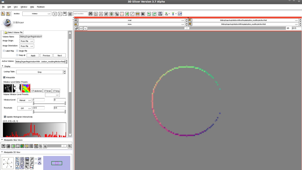
Translated sphere - motion field following registration WITHOUT regularization: | |||
[[File:translatedSphereMotionFieldWithoutRegularization.png| 1000px]] | |||
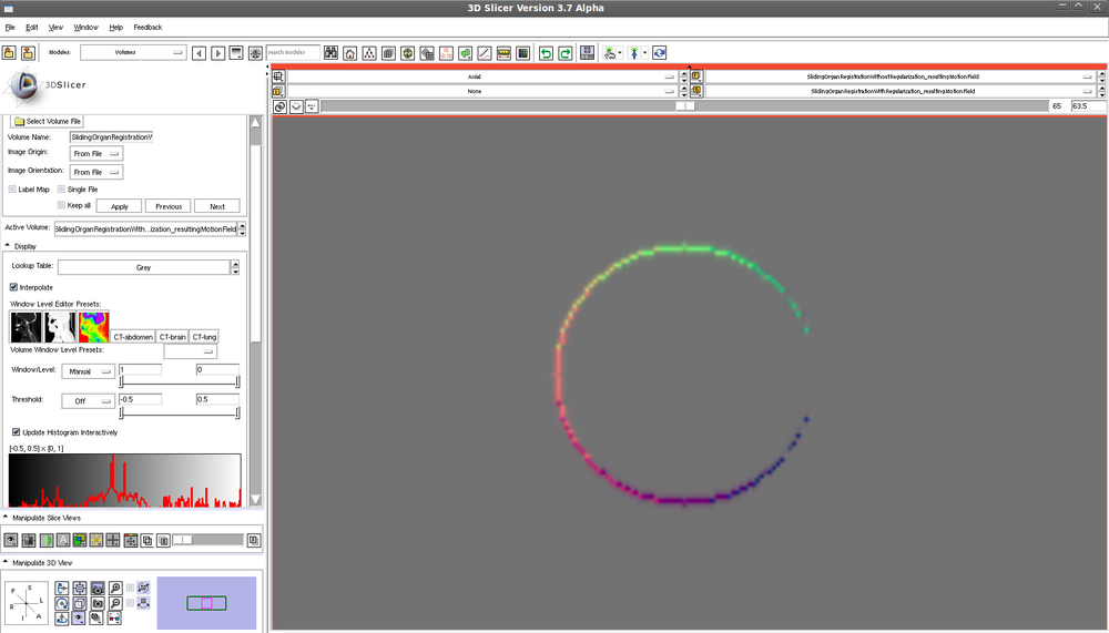
Translated sphere - motion field following registration WITH regularization: | |||
[[File:translatedSphereMotionFieldWithRegularization.png| 1000px]] | |||
== XCAT phantom == | == XCAT phantom == | ||
| Line 43: | Line 51: | ||
*** for index in range - generate spherical lesion at index, use nurbs_save = 1 to save lesion.nrb, then calculate centroid of lesion control points | *** for index in range - generate spherical lesion at index, use nurbs_save = 1 to save lesion.nrb, then calculate centroid of lesion control points | ||
[[File: | [[File:2010_10_19_motionFieldParaviewOverview.png|800 px]] | ||
** --segmentation (XCAT mode | *** Question: are there discontinuities / sliding motion. Looks like it =) See circled region: | ||
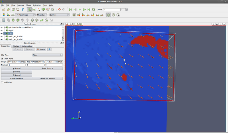
[[File:2010_10_19_goldStandardMotionFieldDiscontinuities_annotated.png|800 px]] | |||
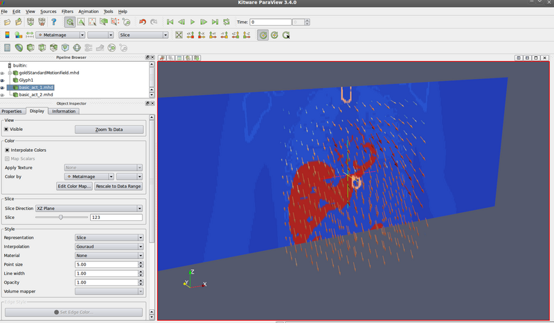
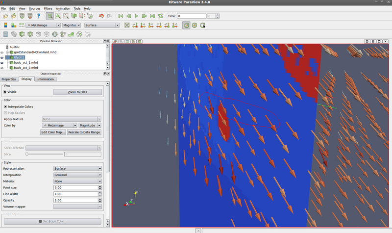
*** Question: does the motion field make sense? Yes: | |||
Fixed image (look at origin of vectors): | |||
[[File:2010_10_19_motionFieldParaview_1.png|800 px]] | |||
Moving image (look at destination of vectors): | |||
[[File:2010_10_19_motionFieldParaview_2.png|800 px]] | |||
** --segmentation (XCAT mode 5 - save anatomical variation mode, or run XCAT mode 0 with save_nurbs = 1) to save surfaces (.nrb text file). Then create new .txt file for the surface you want. Then run itkImageToImageDiffusiveDeformableRegistrationGenerateNormalsTest to create itk::Image<PixelType, Dimension> of control points | |||
*** Unfortunately: (1) control points need interpolation, can't find NURBS support in VTK; (2) XCAT has MANY types of surfaces for one organ - ex all intracardiac surfaces, etc, and it would be a lot of work to sort through them; (3) Perfect segmentation not great for registration method evaluation - removes segmentation error component of total error. | *** Unfortunately: (1) control points need interpolation, can't find NURBS support in VTK; (2) XCAT has MANY types of surfaces for one organ - ex all intracardiac surfaces, etc, and it would be a lot of work to sort through them; (3) Perfect segmentation not great for registration method evaluation - removes segmentation error component of total error. | ||
*** Ex: right lung | |||
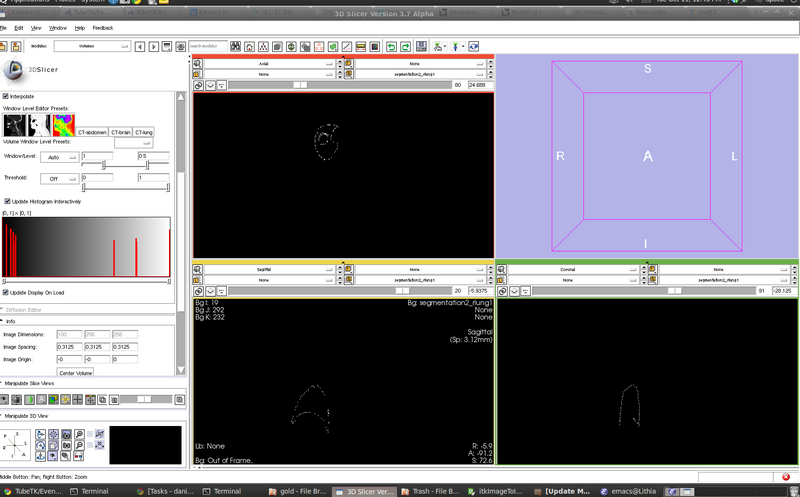
[[File:segmentation2_rlung.png | 800 px]] | |||
== Talking points == | == Talking points == | ||
| Line 56: | Line 79: | ||
* Extract organ border normals from input data | * Extract organ border normals from input data | ||
* Run registration + compare to gold standard for validation #s | * Run registration + compare to gold standard for validation #s | ||
[[Category:TubeTK Events and Meetings|2010.10.19]] | |||
Latest revision as of 18:48, 26 July 2013
- Dashboard: https://www.kitware.com/CDash/index.php?project=TubeTK
- Registration re-write needed
- Wiki Documentation: http://public.kitware.com/Wiki/TubeTK
Stephen
Completed
- Ridge extractor success
- Email requests for TubeTK
- Licensing changes
- Blur3D -> Blur
Ongoing
- Radius extractor tests
- 2D and 3D only :(
- Funding re-allocation
Upcoming
- Application in plant biology
- Proposal with Paul / Liz
Romain
- Implementation on the ComputeTortuosity module (only Distance Metric)
- Started debugging
- Future
- More debugging! ---> hopefully testing soon
- #tube command
- Implementation of the other 2 methods : ICM SOAM
Danielle
Sliding organ registration
- Fixed "duh" problem from Thursday - extracting tangential and normal components before computing regularization
- Regularization smoothing in normal direction completed
- Added intensity distance function (mean squares)
Translated sphere - motion field following registration WITHOUT regularization:
Translated sphere - motion field following registration WITH regularization:
XCAT phantom
- Got XCAT running on my machine (thanks Andinet!)
- Learned XCAT parameters
- Wrote isbiValidation.py:
- --basic (XCAT mode 0 - phantom mode): with respiratory period 5s and cardiac period 1s, generates two images at 0s and 2.5 s, saves as .mhd
- --gold (XCAT mode 2 - spherical lesion generation mode): creates gold standard motion field, and saves as .txt (index, vector in 6 columns). Then run itkImageToImageDiffusiveDeformableRegistrationGenerateGoldStandardTest to create itk::Image<VectorType, Dimension> .mhd for visualization
- XCAT phantom gives gold standard vectors for surface borders (XCAT mode 4), but no internal vectors! So...
- for index in range - generate spherical lesion at index, use nurbs_save = 1 to save lesion.nrb, then calculate centroid of lesion control points
- Question: are there discontinuities / sliding motion. Looks like it =) See circled region:
- Question: does the motion field make sense? Yes:
Fixed image (look at origin of vectors):

Moving image (look at destination of vectors):
- --segmentation (XCAT mode 5 - save anatomical variation mode, or run XCAT mode 0 with save_nurbs = 1) to save surfaces (.nrb text file). Then create new .txt file for the surface you want. Then run itkImageToImageDiffusiveDeformableRegistrationGenerateNormalsTest to create itk::Image<PixelType, Dimension> of control points
- Unfortunately: (1) control points need interpolation, can't find NURBS support in VTK; (2) XCAT has MANY types of surfaces for one organ - ex all intracardiac surfaces, etc, and it would be a lot of work to sort through them; (3) Perfect segmentation not great for registration method evaluation - removes segmentation error component of total error.
- Ex: right lung
- --segmentation (XCAT mode 5 - save anatomical variation mode, or run XCAT mode 0 with save_nurbs = 1) to save surfaces (.nrb text file). Then create new .txt file for the surface you want. Then run itkImageToImageDiffusiveDeformableRegistrationGenerateNormalsTest to create itk::Image<PixelType, Dimension> of control points
Talking points
- Underlying XCAT deformation fields too perfect? They are likely interpolating the border vectors.
Future
- Boundary conditions
- Compute weighting term = distance to organ border
- Extract organ border normals from input data
- Run registration + compare to gold standard for validation #s
